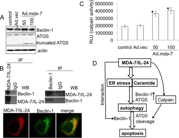
Fig. 6. Role of Beclin-1 and ATG-5 in Ad.mda-7-induced apoptosis.
A, Expression profile of Beclin-1 and ATG5 protein levels 48 h post-infection of DU-145 cells with Ad.mda-7 (50 or 100 pfu/cell) or Ad.vec (100 pfu/cell). B, DU-145 cells were infected with 100 pfu/cell of Ad.vec or Ad.mda-7 for 48 h and immunoprecipitated with anti-MDA-7/IL-24 or anti-Beclin-1 followed by immunoblotting with anti-Beclin-1 or anti-MDA-7/IL-24 antibodies (upper panel). Fluorescent confocal micrographs of DU-145 showing co-immunolocalization of Beclin-1 and MDA-7/IL-24 (lower panel). C, DU-145 cells were infected with Ad.vec (100 pfu/cell) or Ad.mda-7 (50 or 100 pfu/cell) for 48 h followed by calpain assay using calpain-Glo assay. Values reported are mean ± S.D. of three independent experiments. *, p < 0.05 versus control cells. D, Model illustrating the possible molecular mechanism of ER stress- and ceramide-mediated autophagy promoted by MDA-7/IL-24 that switches to apoptosis in prostate cancer cells.

