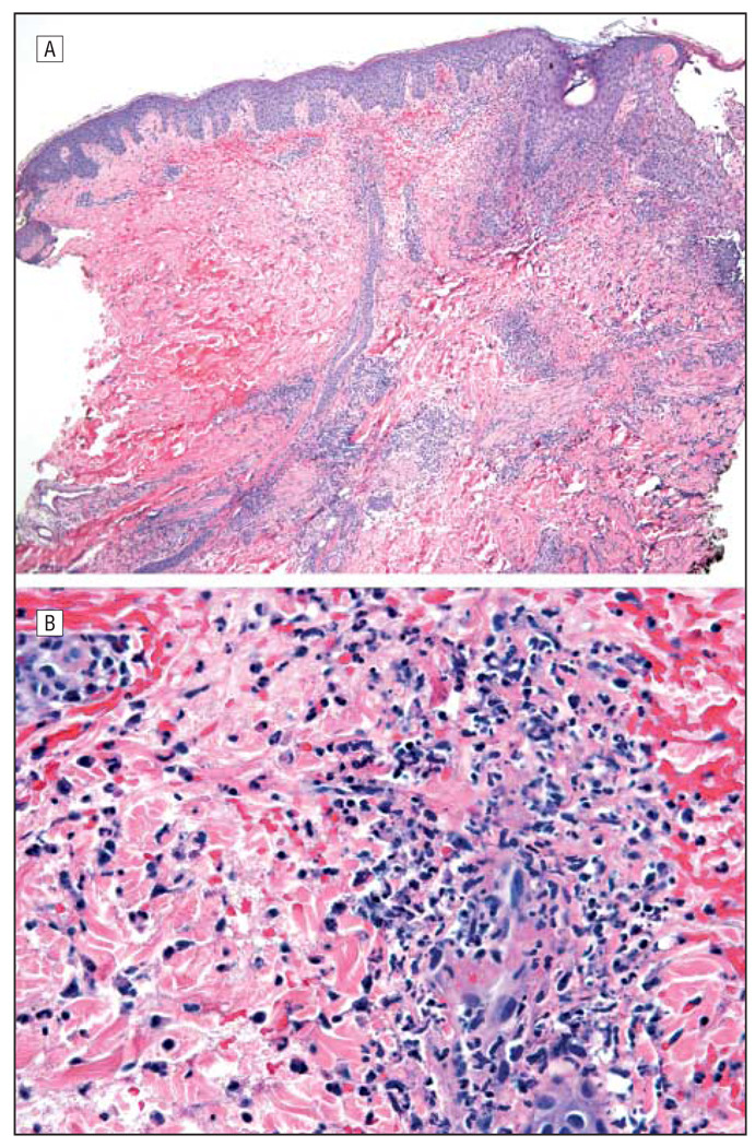
Figure 2.
Histopathologic images from our case. A, Low-power hematoxylin-eosin–stained specimen of a skin lesion showing peripheral epidermal blister formation and a dense dermal infiltrate with leukocytoclasia but no evidence of vasculitis (original magnification ×40). B, High-power hematoxylin-eosin–stained specimen reveals an infiltrate composed largely of neutrophils (original magnification ×400). Findings under Gram, Grocott, and acid-fast bacilli stains were all negative.

