Figure 6.
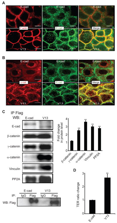
Transfection of V13 into A253 cancer cells enhances intercellular adhesion. (A), Immunofluorescence localization of E-cadherin and F-actin in A253 cells following transfection with either E-cad or V13. E-cadherin from V13 cells exhibit increased co-localization with F-actin at cell-cell contact sites. E-cad and V13 cells were grown to confluence and processed for indirect immunofluorescence staining using an antibody to the cytoplasmic region of E-cadherin. Cells were counterstained for F-actin with rhodamine-phalloidin. Shown are 0.74 μm confocal x–y sections. Size bars, 10 μm. (B) Immunofluorescence localization of E-cadherin and α-catenin in A253 cells transfected with E-cad and V13. Enhanced lateral colocalization of E-cadherin and α-catenin was detected in V13 cells compared to E-cad cells. Shown are 0.74 μm confocal x–y section. Size bars, 10 μm (C) A253 cells transfected with V13 remodel AJs with increased amounts of stabilizing proteins. E-cadherin complexes were immunoprecipitated from A253 cells transfected with E-cad and V13 using Flag antibody. Samples were analyzed for association with β-catenin, γ-catenin, α-catenin, vinculin and PP2A by WB. Bargraph, Fold changes in protein levels associated with E-cadherin from V13 cells were determined in comparison to E-cad after normalization to Flag (*P < 0.05). (D) Hypoglycosylated E-cadherin variant, V13 enhances TER in A253 cells. The TER for nontransfected A253 cells was defined as 1.0. Error bars reflect standard deviation from three independent studies; P values were calculated by two-tailed t-test.

