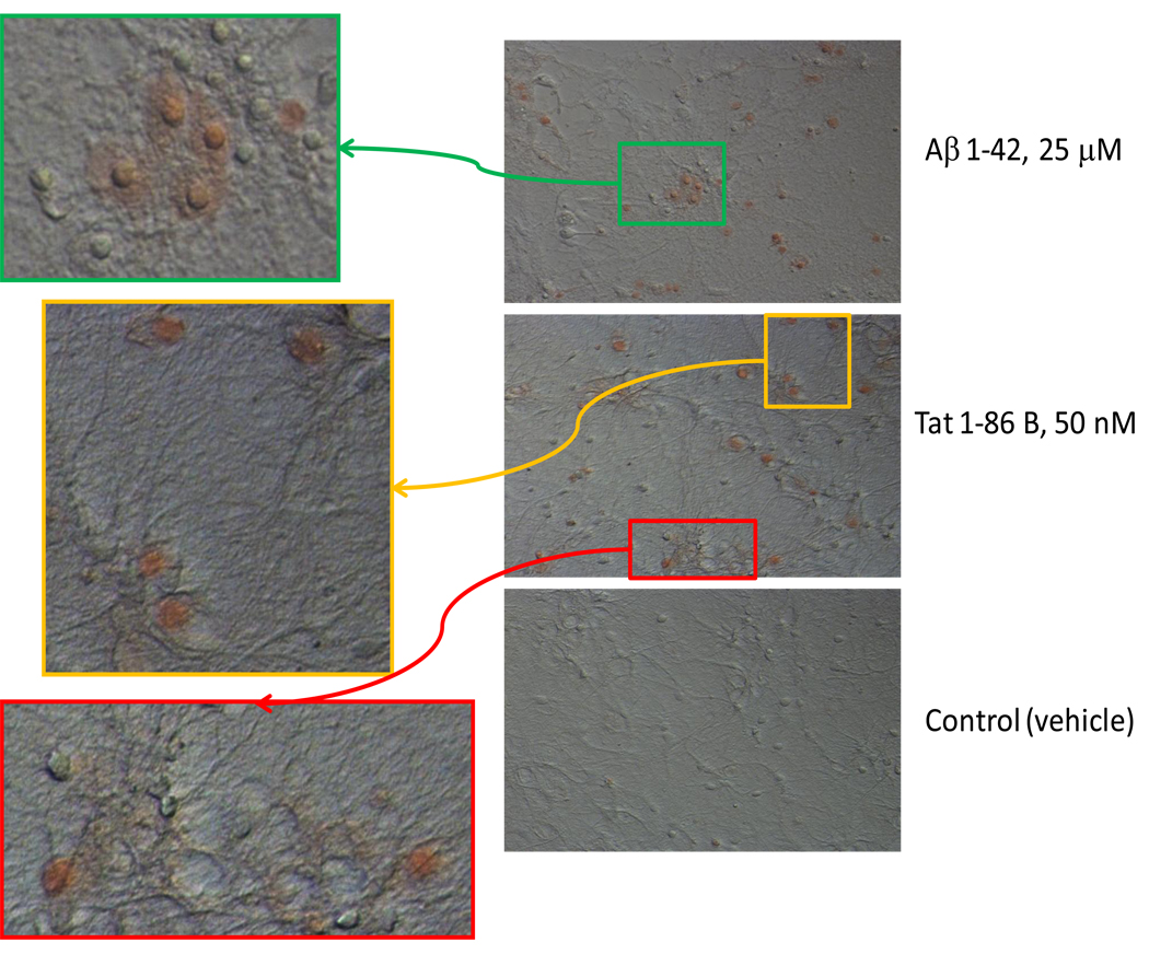
Figure 4. The detection of cell-bound β-amyloid aggregates by the Congo Red staining in living hippocampal cells exposed to Aβ 1–42 or HIV-1 Tat 1–86 B.
Representative images show the Congo Red binding to hippocampal cell cultures exposed to a toxic dose of Aβ 1–42 (40-hour exposure), Tat 1–86 B (24-hour exposure), or vehicle. Results of the Congo Red staining in cultures treated with 100 nM gp120 for 72 hours, 100 nM Cys22 Tat or Tat 1–101 C for 24 hours were not different from the vehicle-treated controls and are not shown in the Figure. Colored boxes mark areas of Congo Red/DIC images, which are shown as new magnified images with greater resolution.

