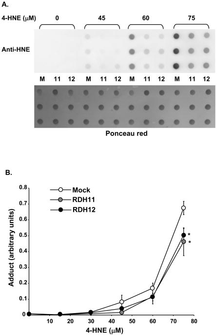
Figure 3.
Mouse RDH11 and RDH12 protect against 4-HNE-protein adduct formation in HEK-293 cells. Confluent cells were treated overnight (20 h) with indicated concentrations of 4-HNE. A, Whole cell homogenates are prepared and equal aliquots (5 μg) of protein are analyzed by dot blot. M, mock; 11, RDH11; 12, RDH12. Protein loading is first verified by staining the membrane with Ponceau red (lower picture) and the membrane is then incubated with anti-HNE coupled with HRP at 1:1000 dilution. B, Graph shows quantification of adduct formation in 6 independent experiments. Data points represent the mean and error bars denote SEM. RDH-expressing clones were compared with the control using the Student’s t test for significance. *=p<0.05; **=p<0.001; and ***=p<0.0001.

