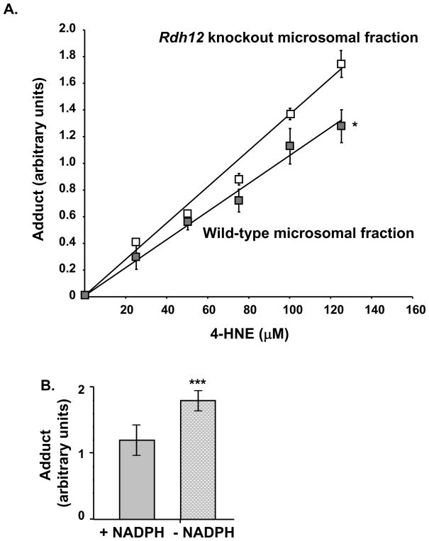
Figure 4.
RDH12 protects against formation of 4-HNE-protein adducts in mouse retinal microsomes. Microsomal fractions were prepared from wild-type and Rdh12 knockout retinas. A, Equal aliquots (10 μg) of microsomal proteins from wild-type or Rdh12 knockout retinas were incubated with indicated concentrations of 4-HNE in reaction buffer containing NADPH, for 2 h at room temperature. Reactions were transferred to the membrane by vacuum filtration, and unbound 4-HNE was washed 3 times with PBS. Protein loading is verified and adduct formation is quantified as described in Figure 3. Graph shows quantification of adduct formation in 3 independent experiments. B, The same experiment is repeated with wild-type retinal microsomes and 125 μM of 4-HNE, with and without NADPH. Graph shows quantification of adduct formation in 3 independent experiments. Data points represent the mean and error bars denote SEM. Results were compared using the Student’s t test for significance. *=p<0.05; **=p<0.001; and ***=p<0.0001.

