Figure 2.
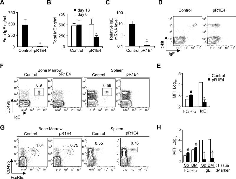
Effect of in vivo expression of secreted form of chimeric single chain anti-IgE on markers of IgE expression. Two month old BALB/c mice were given pR1E4 or control plasmid i.v. and analyzed 13 days later.
(A) Free serum IgE level measured on d13 after plasmid treatment.
(B) Total serum IgE concentration before treatment (open bars) and 13 days after treatment (filled bars).
(C) qPCR comparison of relative IgE mRNA levels in the spleen of control or pR1E4-treated mice at d13 of treatment.
(D,E) Flow cytometry analysis of IgE bound to peritoneal mast cells in control and pR1E4-treated mice. Peritoneal mast cells were defined as c-kit+FcεRI+CD4-CD8-B220- cells. Mean fluorescence intensity (MFI) of binding by FcεRIα and anti-IgE antibodies was monitored.
(F,G) Analysis of IgE bound to basophils in the spleen and bone marrow of control or treated individual mice. Basophils were identified as CD49b+FcεRI+CD4-CD8-B220-.
(H) Levels of surface IgE and FcεRI on basophils in the spleen and bone marrow on day 13 post plasmid treatment. Results are means ± s.d. of 8 mice receiving pR1E4 compared to 5 mice receiving empty vector. Statistical significance of differences between pR1E4-treated and control-treated mice was calculated using Student's T test: *P < .001; #P < 0.2.

