FIGURE 3.
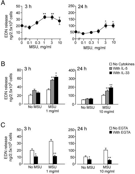
MSU crystals induce modest degranulation of eosinophils, which is regulated by cytokines and extracellular calcium. (A) Eosinophils were incubated with MSU crystal suspensions for 3 h or 24 h at 37 °C. EDN released into supernatants was measured by ELISA. Results show the mean±SEM from eight (for 3 h) or four (for 24 h) different eosinophil preparations. * and **; significant differences compared with medium alone (p<0.05 and p<0.01, respectively). (B) Eosinophils were incubated with or without 1 mg/ml (for 3 h) or 10 mg/ml (for 24 h) MSU crystal suspensions at 37 °C with or without 1 ng/ml IL-33 or 1 ng/ml IL-5. EDN released into supernatants was measured by ELISA. Results show the mean±SEM from eight (for 3 h) or four (for 24 h) different eosinophil preparations. * and **; significant differences compared with the samples with MSU but no cytokines (p<0.05 and p<0.01, respectively). (C) Eosinophils were incubated with or without 1 mg/ml (for 3 h) or 10 mg/ml (for 24 h) MSU crystal suspensions at 37 °C with or without 1 mM EGTA. EDN released into supernatants was measured by ELISA. Results show the mean±SEM from eight (for 3 h) or four (for 24 h) different eosinophil preparations. * and **; significant differences compared with the samples without EGTA (p<0.05 and p<0.01, respectively).

