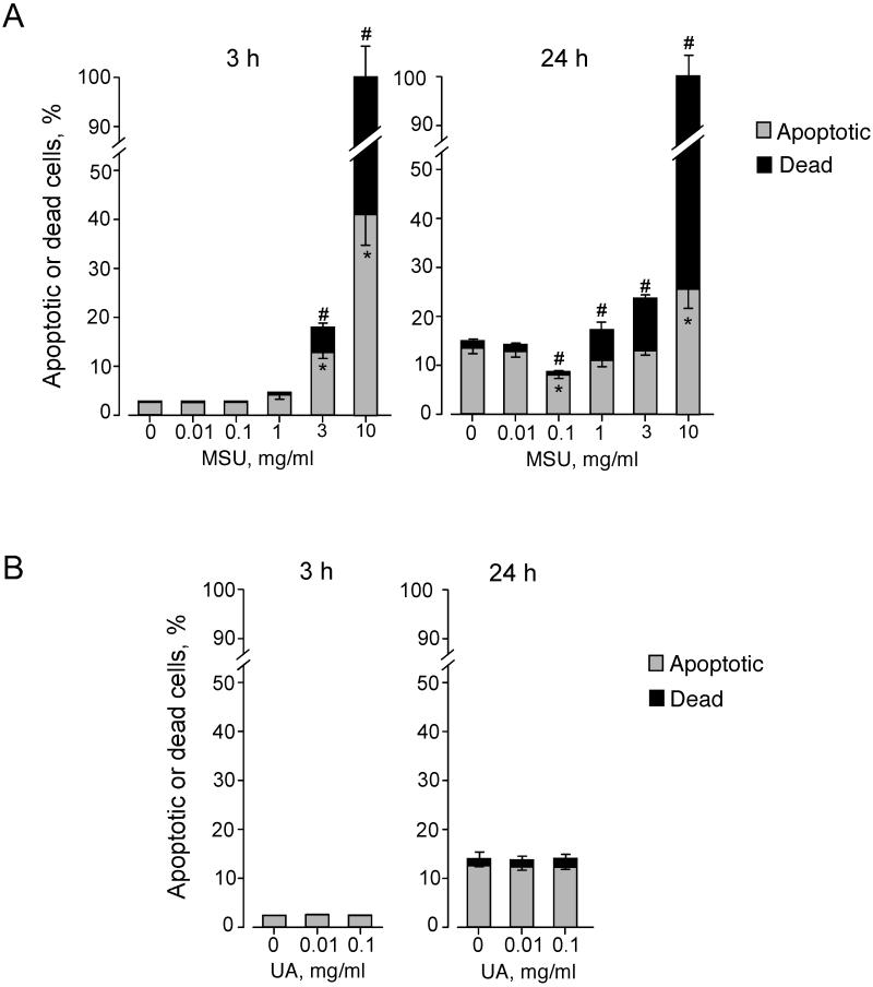
FIGURE 4.
MSU crystals modulate eosinophil viability. (A) Eosinophils were incubated with medium alone or serial dilutions of MSU crystal suspensions for 3 h or 24 h at 37 °C. (B) Eosinophils were incubated with medium alone or serial dilutions of soluble uric acid (UA) for 3 h or 24 h at 37 °C. Cell apoptosis and death were examined by double staining with FITC-conjugated annexin V and PI and flow cytometry analysis. The percentages of apoptotic cells (annexin V-positive and PI-negative) and dead cells (annexin V-positive and PI-positive) were determined. The proportion of cells with annexin V-negative and PI-positive was less than 0.5%. Results show the mean±SEM from five (A) or three (B) different eosinophil preparations. Downward error bars and upward error bars indicate SEM for apoptotic cells and dead cells, respectively. * and #; significant differences compared with percent apoptotic cells and dead cells in medium alone, respectively (p<0.01).

