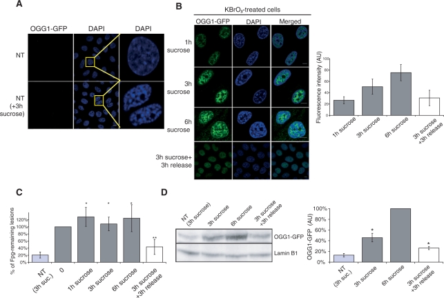
Figure 6.
Chromatin condensation by hypertonic shock impedes 8-oxoG repair. (A) NT cells pre-incubated or not with 250 mM sucrose for 3 h and washed with CSK–0,5% triton buffer prior to fixation. DNA is stained with DAPI. (B) OGG1–GFP cells are treated with 40 mM KBrO3 for 30 min and allowed to recover in DMEM supplemented with 250 mM sucrose. In the last row of images, sucrose was removed after 3 h and replaced by fresh medium for 3 h. Cells were washed with CSK buffer prior to fixation. Graph represents GFP intensity for each condition (number of nucleus > 10). Scale bar = 10 µm. (C) 8-OxoG quantification by alkaline elution of cells treated with 40 mM KBrO3 and recovered in 250 mM sucrose supplemented medium. Bilateral Student test (*P > 0.1 compared to the point 0; **P< 0.04 and P< 0.02 compared to the points 0 and 3 h sucrose respectively). (D) western blot of cells treated with 40 mM KBrO3 and recovered in 250 mM sucrose supplemented medium. Antibodies against GFP are used and lamin B1 is used as a loading control. Quantification of signals using Scan GBox is represented on the graph on the right. Bilateral Student’s test (*P < 0.05).

