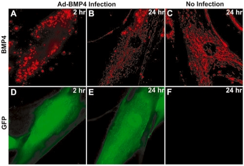
Figure 8.
Fluorescence images of Cy3-labeled BMP-4 targeting MBs in HDF cells that infected with (A, B, D, and E) and without (C and F) adenovirus. Infected cells highly expressed BMP-4 and GFP, which was used as an independent marker for infection, but not tagged to BMP-4 protein. HDF cells were infected with adenovirus and then delivered with 1 µM of BMP-4 targeting MB. MB (A–C) and GFP (D–F) fluorescent signals were observed 2 (A and D) and 24 (C and E) hours after delivery. The same exposure time was used for each channel.

