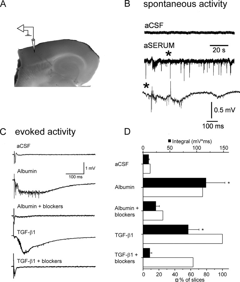
Figure 1.
TGF-β signaling induces epileptiform activity. A, Photograph of a brain slice displaying electrode positioning. The stimulating electrode was placed at the white–gray matter border. B, Extracellular recordings showing spontaneous interictal-like epileptiform activity after treatment with aSERUM containing albumin. Asterisk refers to the region corresponding to the slower time scale shown in the lower trace. C, Evoked responses from slices treated with aCSF, albumin, albumin plus TGF-β receptor blockers, TGF-β1, or TGF-β1 plus TGF-β receptor blockers. TGF-β receptor blockers prevent epileptiform activity induced by albumin or TGF-β1 treatment. D, Comparison of mean event integral (black bars) in the 50–500 ms time range (after stimulation) shows a significant increase in the integral of the delayed epileptiform field potential in the albumin and TGF-β1-treated slices but not in slices treated with TGF-β receptor blockers. The white bars represent the percentage of slices with paroxysmal, epileptiform activity. Error bars indicate SEM. Asterisks indicate p < 0.05.

