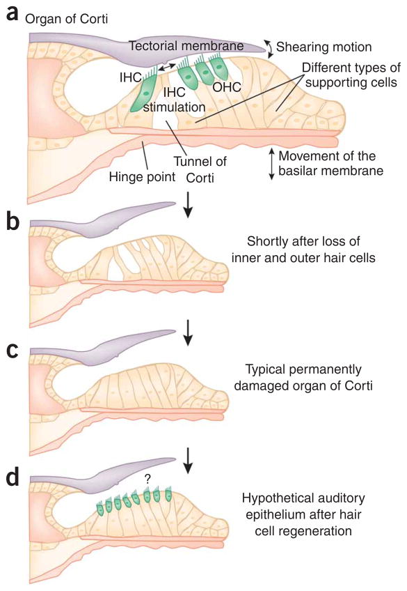
Figure 1.
Conceptual drawings of the normal, damaged and repaired organ of Corti. (a) In the normal organ of Corti, movements of the basilar membrane are relayed by means of hinging near the tunnel of Corti and shearing motions between outer hair cells (OHCs) and the tectorial membrane. The connections of OHC stereocilia with the tectorial membrane are essential for proper function of the cochlear amplifier. All these parts of the organ of Corti must be in appropriate mechanical correlation with one another to ensure proper stimulation of the inner hair cells (IHCs). (b) Complete loss of IHCs and OHCs, shortly after a toxic insult. (c) Collapse of the tunnel of Corti and dedifferentiation of the supporting cells. (d) Hypothetical reseeding of generic hair cells in the damaged organ of Corti epithelium. It is unclear whether a connection with the tectorial membrane will re-form, how far it will stretch over the new hair cell area and whether the hair cells will be appropriately stimulated by movements of the basilar membrane.

