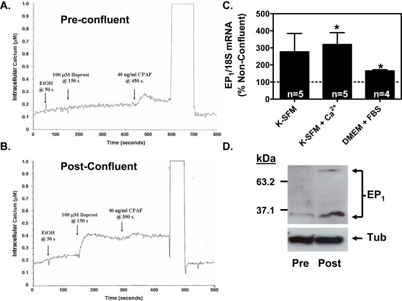
Figure 2. EP1 receptor signaling and expression are up-regulated in PHKs following the attainment of a confluent monolayer.
(A & B). Agonist induced calcium mobilization is restricted to PHKs grown to a post-confluent monolayer. Cells at low cell density (pre-confluent) or high cell density (post-confluent) were loaded with the fluorescent calcium indicator, Fura PE3/AM. After trypsinization, the cells were stimulated with ethanol (EtOH), 100 μM iloprost in ethanol, or 40 ng/ml of the positive control platelet activating factor receptor (PAF-R) agonist, carbamyl-PAF (CPAF). (A). The EP1 receptor agonist, iloprost, is unable to induce measurable calcium mobilization in pre-confluent PHKs. In contrast, the positive control PAF-R agonist, CPAF, is shown to induce a calcium mobilization response. (B). The EP1 receptor agonist, iloprost, induces a robust calcium mobilization response in post-confluent keratinocytes. (C). EP1 receptor mRNA expression is up-regulated in post-confluent PHKs compared with pre-confluent PHKs under differing culture conditions. RNA was prepared from both pre-confluent and post-confluent cells and EP1 expression was assessed by quantitative real-time RT-PCR and normalized to 18S rRNA. Results represent the mean ± SEM for n=4-5 experiments done in duplicate; * p < 0.05, one-sample t-test relative to 100 % for pre-confluent controls. (D). EP1 receptor protein expression is up-regulated in post-confluent PHKs compared with pre-confluent PHKs. An immunoblot was performed on total cell lysates from pre-confluent PHKs and post-confluent PHKs grown in serum free media (K-SFM; 0.06 mM Ca2+). EP1 immunoreactive bands are seen at approximately 35 kDa and 70 kDa. The blot was stripped and reprobed with anti-α-tubulin antibody as a loading control (bottom panel).

