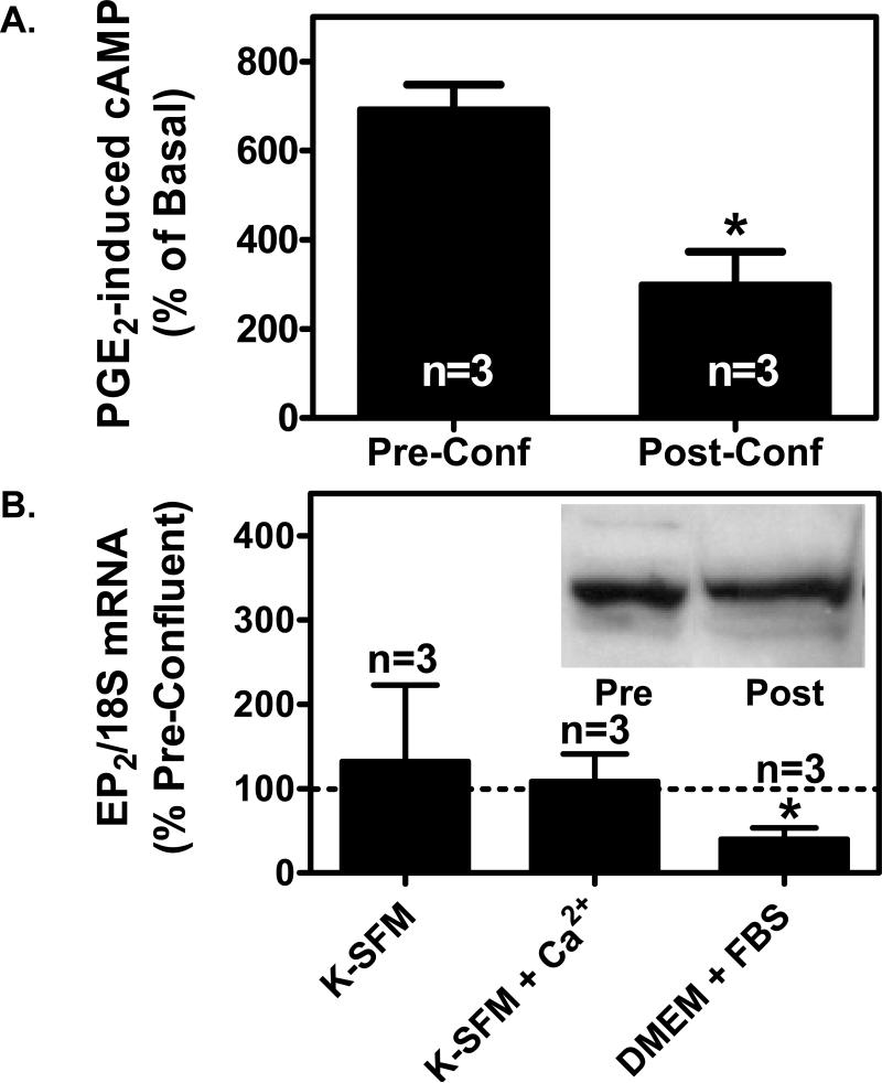
Figure 3. Depending on the culture media, high cell density either has no effect on EP2 receptor expression or suppresses EP2 receptor expression in PHKs.
(A). PGE2-induced cyclic AMP production is reduced in PHKs at high cell density. Pre-confluent PHKs or post-confluent PHKs cultured in DMEM + 10% FBS were treated with 3 μg/ml indomethacin overnight to block endogenous PGE2 formation. The cells were then stimulated with 100 nM PGE2 for 1 minute. Cyclic AMP was measured using a commercial EIA kit as described in the methods section. The results represent the mean and SEM of three experiments. (B). Quantitative real-time PCR was performed for EP2 receptor mRNA expression as described for the EP1 receptor in figure 2C above. Inset: Immunoblot for EP2 receptor expression in pre-confluent (Pre) and post-confluent (Post) PHKs grown in DMEM with 10% fetal bovine serum. Membrane preparations were produced from pre-confluent and 2 days post-confluent PHKs. In each case, 40 μg of the membrane preparation was separated by SDS-PAGE electrophoresis and immunoblot performed using a polyclonal anti-EP2 receptor antibody as described in the methods section.

