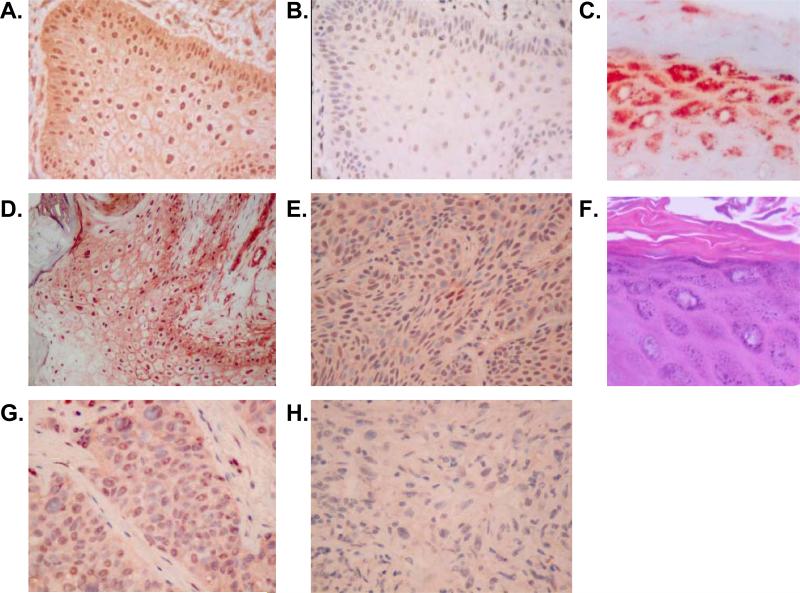
Figure 5. EP1 receptor immunolocalization in non-melanoma skin cancer.
Immunohisochemical (IHC) analyis of EP1 receptor expression was performed on formalin-fixed, paraffin-embedded archival tissue samples. In each case, 5 μm sections were deparaffinized and heat-induced epitope retrieval was done as outlined in the methods section. IHC staining was done using a monoclonal anti-human EP1 receptor antibody (clone 5F12) (A, C-F) or an isotype control (B). (A). Keratoacanthoma (400x magnification). (B). A serial section of the same keratoacanthoma stained with isotype (IgG2bκ) negative control antibody (400x). (C). Hyperplastic skin overlying the basal cell carcinoma seen in panel E. (D). Well-differentiated squamous cell carcinoma (SCC) (200x). (E). Basal cell carcinoma (400x). (F). Hematoxylin & eosin stained section corresponding to the section seen in panel C. (G). Poorly-differentiated SCC (400x). (H). Spindle cell carcinoma (400x).

