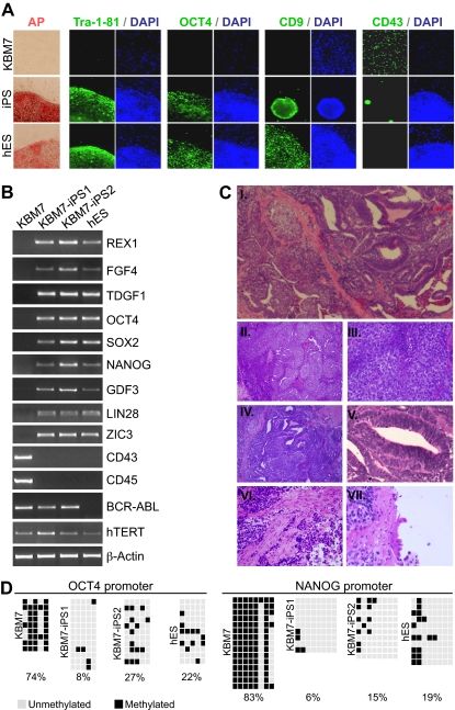
Figure 1.
Generation of iPSCs derived from human KBM7 leukemic cancer cells. (A) Introduction of c-MYC, OCT4, SOX2, and KLF4 into KBM7 cells led to the formation of rare adherent colonies that displayed morphologic similarity to human embryonic stem (ES) cells as well as high alkaline phosphatase activity. Reprogrammed KBM7 cells homogeneously stained for pluripotency markers Tra-1-81 and OCT4 and CD9 but did not stain for hematopoietic CD43 (green immunofluorescence; nuclear counterstain in blue). (B) Total RNA from KBM7 cells, reprogrammed KBM7 cells, and human ES cells was isolated and analyzed by reverse-transcribed polymerase chain reaction for the expression of human ES cell characteristic transcripts REX1, FGF4, and TDGF1, endogenous OCT4 and SOX2, NANOG, GDF3, LIN28, and ZIC3, the hematopoietic specific transcripts CD43 and CD45, and the CML specific BCR-ABL fusion transcript. (C) Subcutaneous injection of cancer induced pluripotent stem cells (iPSCs; KBM7-iPS2) into non-obese diabetic severe combined immunodeficiency (NOD-SCID) mice led to formation of teratoma. Hematoxylin and eosin-stained sections of the tumor (i, original magnification ×40) revealed extensive areas of embryonal carcinoma (ii, original magnification ×100; iii, original magnification ×400) and ectoderm-derived primitive neuroectodermal tissue (iv, original magnification ×100; v, original magnification ×400). Differentiation into muscle (mesodermal; vi, original magnification ×400), and ciliated respiratory epithelium (endodermal; vii, original magnification ×600) was also present. (D) Methylation analysis of the OCT4 and NANOG promoter region in KBM7 cells, the generated iPSCs, and hES cells. Light gray squares represent unmethylated; black squares, methylated cytosine-phosphate-guanosine. Numbers indicate the percentage methylated cytosine-phosphate-guanosine.

