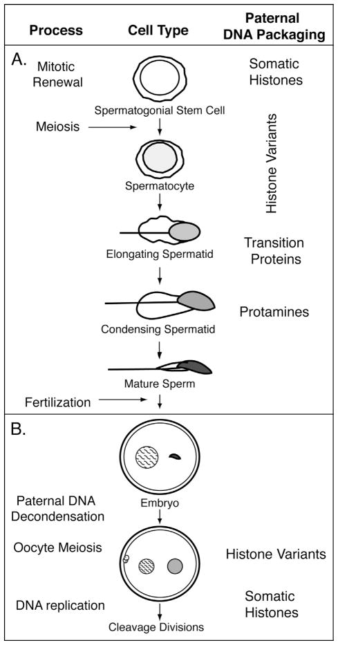
Figure 1.
Schematic diagram of the progression of mammalian sperm cell nuclei through spermatogenesis and early post-fertilization stages. (A) In the testis, proliferating spermatogonial stem cells give rise to mature spermatozoa. During spermatogenesis, the chromatin of developing spermatozoa becomes successively more compact, with accompanying shifts in global chromosomal packaging by sperm nuclear basic proteins. This process is initiated during meiosis, with the bulk of the transition occurring in post-meiotic stages. Concurrently, epigenetic processes demarcate portions of the genome that undergo differential processing during post-meiotic stages (spermiogenesis) or post-fertilization. Developmental processes (stages) are indicated in the left column. Stage-specific incorporation of generalized relevant proteins that change nucleosomal composition are shown on the right. (B) Paternal chromatin (smooth shading) carries epigenetic marks established by chromatin factors during spermatogenesis that make it distinct from maternal chromatin (hatched shading) after fertilization and before the first cell division. Fertilization induces the oocyte to complete meiosis, resulting in one haploid maternal pronucleus and two extruded polar bodies. During this time, paternal chromatin delivered into the oocyte goes through extensive changes including the repackaging of sperm chromosomes with somatic histones, global decondensation, and imprinted transcriptional regulation, but maintains distinct epigenetic features.

