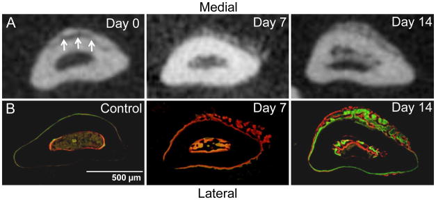
Figure 2.
Abundant woven bone formation near the site of non-displaced fracture. Transverse sections of ulnae (0–1 mm distal to mid-shaft) obtained by microCT (A) and fluorescent microscopy (B). The fracture is seen on the medial side of the ulna on day 0 microCT (arrows). Periosteal and endosteal woven bone is present at low density on day 7 and at higher density on day 14. The periosteal woven bone is thicker medially, corresponding to the fracture location. (Calcein green and alizarin complexone [red] were administered 7 and 2 days prior to sacrifice, respectively.)

