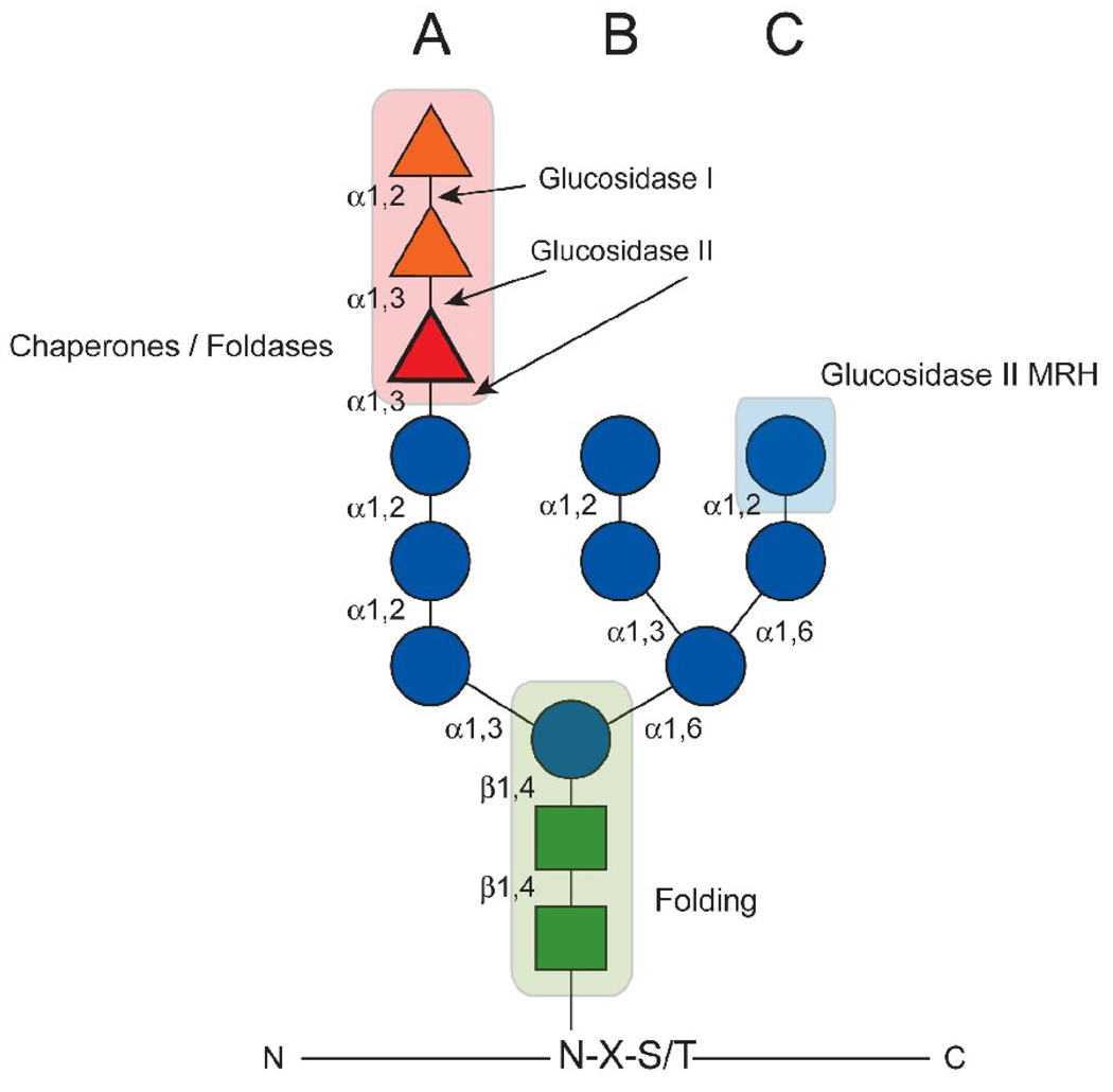
Figure 1.
The composition and roles of N-linked glycans in the ER. The 14-member carbohydrate is covalently linked to Asn residues of the consensus sequence Asn-X-Ser/Thr as depicted. The glycan is comprised of 3 glucoses (triangles), 9 mannoses (circles), and 2 N-acetlyglucosamines (squares). The mannoses are arranged in three branches A, B, and C. The orientations of the glycosidic bonds are indicated. The positions at which glucosidases I and II cleave the glucoses are designated. The glucose that is transferred through GT1 activity and is involved in lectin chaperone binding is highlighted. The regions of the N-glycan that are critical for chaperone interaction, glucosidase II MRH binding, and folding kinetics are shaded.

