
Figure 1.
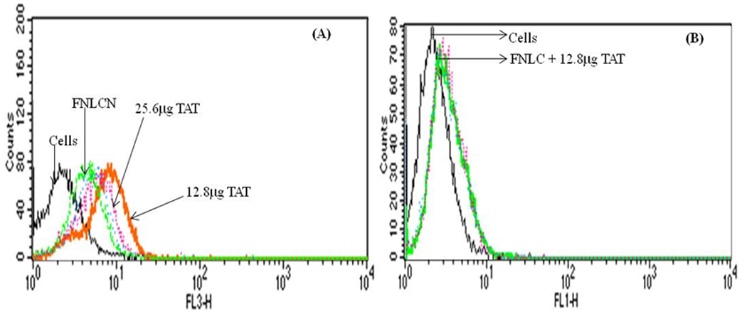
Binding assay of NLC nanoparticles with and without TAT peptide in H460 cell lines. The amount of TAT peptide was optimized with respect to DOGS-NTA-Ni concentration. A) Binding of FNLCN with various amounts of TAT peptide. Increase in TAT peptide concentration above 12.8 µg to 25.6 µg decreased the binding (line is not clear because mixed with blue line of 6.4 µg). B) FNLC formulations without DOGS-NTA-Ni prepared with Dio-dye, incubated with 12.8 µg TAT peptide showed very small non-specific binding.