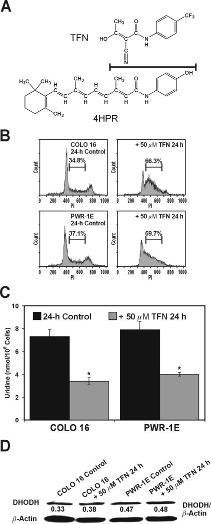
Figure 2.
TFN suppresses DHODH activity in premalignant and malignant human skin and prostate epithelial cells. (A) The chemical structures for TFN and 4HPR. The line separating these structures also designates their chemical similarity. (B) Representative PI histograms showing the percent S phase cells for the COLO 16 and PWR-1E cells exposed for 24 h to Me2SO (control) or 50 μM TFN. (C) The cellular uridine levels were determined for ~106 cells exposed to Me2SO (control) or 50 μM TFN for 24 h. *P<0.001 compared to the respective controls. (D) An immunoblot analysis of DHODH expression for the COLO 16 and PWR-1E cells exposed to Me2SO (control) or 50 μM TFN for 24 h.

