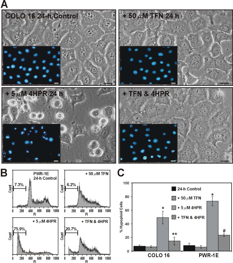
Figure 4.
TFN inhibits 4HPR-induced apoptosis in COLO 16 and PWR-1E cells. (A) Micrographs showing COLO 16 cells exposed to Me2SO (control), 50 μM TFN, 5 μM 4HPR, or 50 μM TFN and 5 μM 4HPR for 24 h. The insets images illustrate the nuclear morphology of the COLO 16 cells as determined by Hoechst 33342 staining and epifluorescence microscopy. The scale bars equal 18 μm. B, Representative PI histograms showing the percent hypoploid cells for the PWR-1E cells treated as described above in (A). The gated cells detected below ~300 fluorescence units of PI on the linear x-axis of the representative histograms are designated the hypoploid apoptotic cell population. (C) A summary of the hypoploid cells in the treatment populations described in (A) and (B). *P<0.001 compared to the respective controls, **P<0.001 compared to the COLO 16 4HPR treatment, and #P<0.01 compared to the PWR-1E 4HPR treatment.

