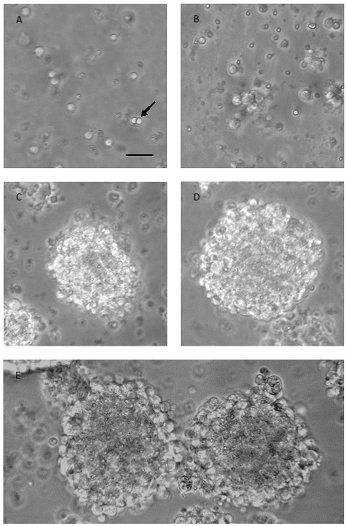
Figure 2. E10 cells gradually form neurospheres.
Phase-bright micrographs showing progressive growth of cells into clusters during the first days in culture with EGF and FGF2. (A) E10 cells at plating day (day 1) are separated into single cells. (B) E10 cells on day 2 have formed clusters of 5–15 cells. (C) E10 cells at day 3 show the formation of bigger, non-rounded clusters. (D) E10 cells at day 4, after being re-plated on day 3, form bigger and rounder clusters. (E) E10 cells at day 5 have grown into round neurospheres. Scale bar 50µm; applies A–E.

