Fig. 3.
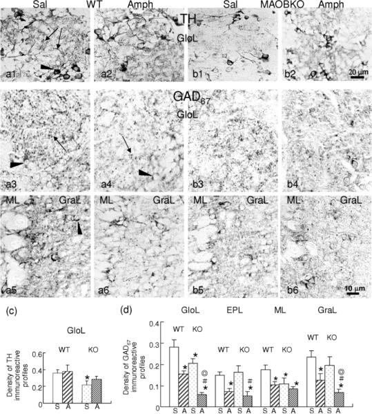
Amph induces differential changes in the expression of tyrosine hydroxylase (TH) or GAD67 in the distinct layers of main olfactory bulb (OlfB) of WT and MAOB KO mice. Representative micrographs (a–b) and bar graphs (c, d) illustrate that the level of TH is higher in the OlfB glomerular layer (GloL) of the WT mouse (a1, a2) than that of the KO (b1, b2) after repeated saline injections (S), and that the levels of GAD67 are reduced to different extents in respective OlfB layers of WT (a3–a6) and KO (b3–b6) mice by repeated Amph injections. The mice received multiple injections of Amph or saline (Sal), and then were perfused with fixative 4 h later, followed by preparation of coronal paraffin OlfB sections and immunocytochemical analysis using an anti-TH or GAD67 antibody, as described in the Methods. The TH and GAD67 immunoreactivity is seen in the perikarya (arrowheads), processes and punctates (arrows). Levels of the immunoreactive profiles were quantified by using an image analysis computer system. The densities of TH and GAD67 are presented as means ± SDs (c, d). *: significantly different from the value of saline-treated WT mice, p<0.05; ⊚: significantly different from Amph-treated WT; #: significantly different from saline-treated MAOB KO, p<0.05. GloL: glomerular layer; EPL: external plexiform layer; ML: mitral cell layer; GraL: granule cell layer.

