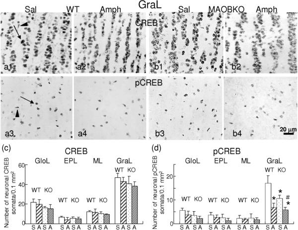
Fig. 4.

Differential expression of CREB and pCREB in OlfB layers of Amph-treated WT and MAOB KO mice. Immunostaing with an anti-CREB or pCREB antibody was performed on coronal OlfB sections of mice at 4 h post-repeated saline or Amph administration. Micrographs of the GraL illustrate that the CREB or pCREB immunoreactivity is seen in the nuclei of neurons (arrows) and glia (arrowheads) of WT (a1–a4) and MAOB KO (b1–b4) mice. *: significantly different from the value of saline-treated WT; p<0.05. #: significantly different from saline-treated MAOB KO. GloL: glomerular layer; EPL: external plexiform layer, ML: mitral cell layer; GraL: granule cell layer.
