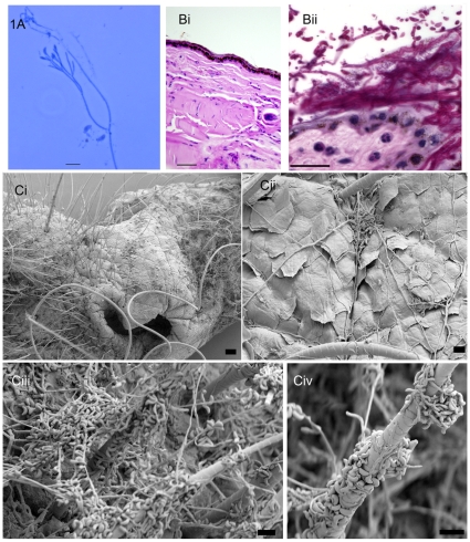
Figure 1. Microscopic and histopathological evidence of G. destructans in bats with WNS.
(A) Direct lactophenol cotton blue mount prepared from skin scrape taken from the muzzle of a little brown bat from Graphite Mine on April 6, 2008 revealed fungal hyphae and curved conidia, bar 10 µm. (B) Control, [Bi] and infected muzzle tissue section [Bii] stained with PAS revealed epidermal colonization by fungal hyphae and spores; the sample was from a little brown bat from Williams Hotel Mine on March 27, 2008. Notably, a few neutrophils are present in the underlying dermis (arrows), bar 10 µm. Bacteria are also seen in this sample (C). SEM photomicrograph of muzzle sample from bat from Williams Hotel Mine showing characteristic curved conidia and septate hyphae spread over bat skin tissues. Note heavy fungal growth with profuse curved conidia covering the skin and hair shaft (Ci, muzzle, bar 100 µm; Cii, higher magnification of a portion of muzzle, bar 10 µm; Ciii & Cvi, higher magnifications, bar 10 µm).

