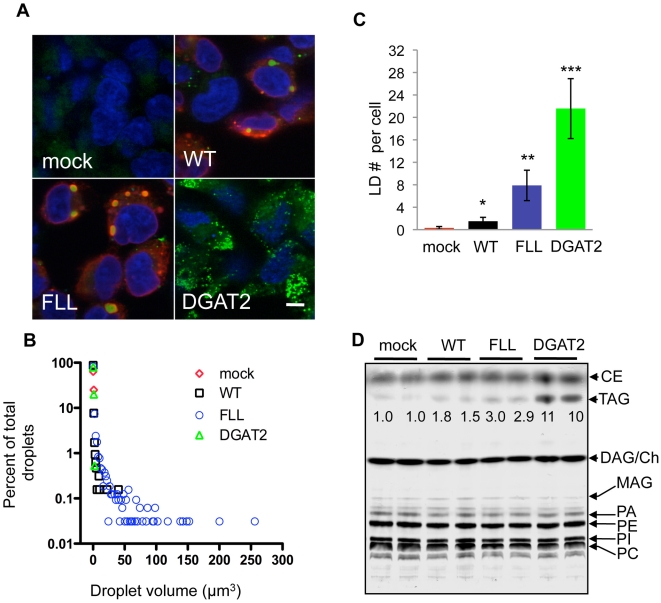
Figure 4. FLL(157-9)AAA FIT2-V5 is a gain-of-function mutant.
A, Confocal fluorescence microscopy of lipid droplets in HEK293 cells transiently transfected with wild-type FIT2-V5 (WT), FLL(157-9)AAA FIT2-V5 (designated FLL), human DGAT2, or mock transfected control. Images are representative of three independent experiments. Cells were stained for lipid droplets (green) and FIT2-V5 (red), and nuclei (blue). B, Histogram of lipid droplet volumes. HEK293 cells were set up as in A and subjected to confocal fluorescence microscopy. Z-stacks were captured and 3D renderings were constructed. Data was analyzed using Velocity software (Perkin Elmer). Analysis was performed on 10 independent 3D renderings from three independent experiments. C, Quantification of lipid droplets per cell is shown (mock vs WT*, FLL**, and DGAT2***: p<3×10⁻⁴, 2.5×10⁻⁵, 4.9×10⁻⁷, respectively). The data are presented as mean ± s.d. D, Quantification of cellular triglyceride concentration in cells expressing the indicated constructs. DGAT2 or mock transfected were used as positive and negative controls, respectively. The data are presented as fold-increase over mock control and are representative of three independent transfections.

