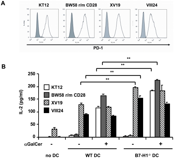
Figure 6. NKT cell hybridoma cells express PD-1 but only the type II NKT cells are stimulated by DC and are negatively regulated by B7-H1.
A) NKT cell hybridoma cells s were stained for PD-1 and analyzed by FACS. The shaded histogram shows the isotype control staining and the black line shows PD-1 staining. B) DC from WT and B7-H1−/− mice were co-cultured with the indicated NKT hybridoma cells in the presence or absence of αGC (10 ng/ml). After 24 hours, the supernatants were tested for IL-2 content by ELISA. Results show 1 out of 4 representative and independent experiments. ** p<0.01.

