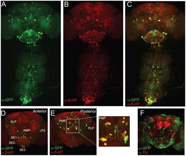
Figure 3. A comparison of TRH-Gal4 driven GFP expression and 5HT immunostaining in male Drosophila brains.
(A–C) TRH-Gal4 (3rd chromosome line) driven mCD8∶GFP signal (A), 5HT immunostaining (B) and overlay (C) of the staining patterns in the brain and the ventral cord of an adult male. (D–F) Anterior (D) and posterior (E) adult male brain 5HT clusters visualized by TRH-Gal4 driven nuclear nls∶GFP (green) expression and 5-HT immunostaining (red). Note that anterior AMP cells are visible with UAS-nls∶GFP (D), but not with UAS-mCD8∶GFP (A). (F) The absence of overlap between TRH-Gal4 driven UAS-nls∶GFP (green) and DA-containing neurons visualized by TH immunostaining (red).

