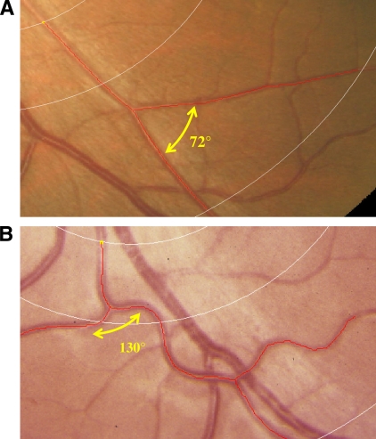
Figure 1.
Measures of retinal vessel tortuosity, branching angle, and optimality deviation. A comparison of optimal (A) and less optimal (B) arrangement of arteriolar geometry. A: Tortuosity of 0.00, branching angle of 72°, and optimality deviation of 0.16. B: Tortuosity of 47.2 × 10⁻³, branching angle of 130°, and optimality deviation of 0.61. A high-quality color representation of this image is available online.

