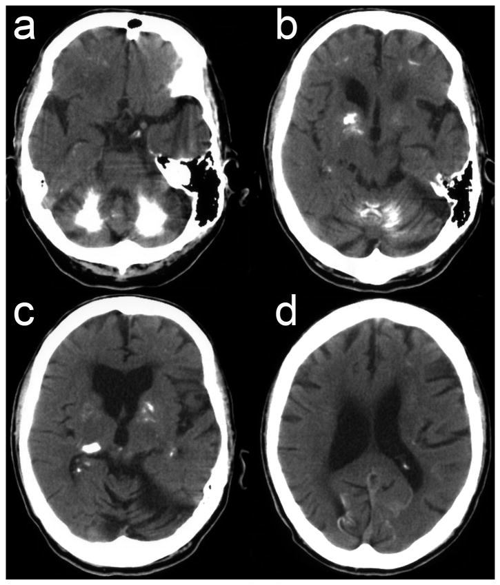
Figure 1.
Brain axial CT-scan shows third and lateral ventricles enlargement and severe calcification of the cerebellar hemispheres and vermis (a,b), striatum, globus pallidus and thalamus (b,c), and subcortical white matter and occipital cortex (a-d). Basal ganglia calcification predominates on the right side (b,c), ipsilateral to symptomatic predominance.

