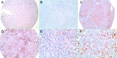
Figure 1.
Examples of immunostaining. (A,D) CD68 stain showing low and high numbers of macrophages, respectively. (B,E) Forkhead box protein staining shows a perifollicular pattern (B). High magnification shows nuclear expression (E). (C,F) A case showing expression of myeloma-associated antigen-1 at low (C) and high (F) magnifications. For all images, low magnification = ×100, whereas high magnification = ×400 (original magnification).

