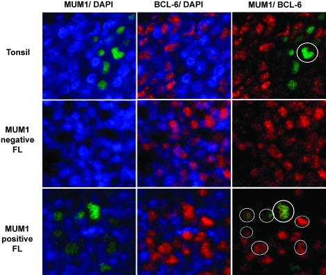
Figure 3.
Dual myeloma-associated antigen-1 (MUM-1)/bcl-6 quantum dot immunofluorescence. Sections were stained with MUM-1 (Qdot 605) and bcl-6 (Qdot655) and counterstained with diamidino-2-phenylindole to highlight nuclei. Images were digitally colorized (MUM-1 green and bcl-6 red). The left panels show MUM-1 alone, middle panel bcl-6 alone, and right panels show combined overlay in the same microscopic field. The top row shows a reactive germinal center from a tonsil in which only rare cells express MUM-1, numerous cells express bcl-6, and only a single cell coexpresses the two proteins (circle). The middle row, from a MUM-1-negative follicular lymphoma (FL), shows malignant FL cells expressing only bcl-6. The bottom row, from a MUM-1-positive FL, shows several MUM-1-expressing cells and numerous bcl-6-expressing cells. The MUM-1 and bcl-6 colocalize (circles).

