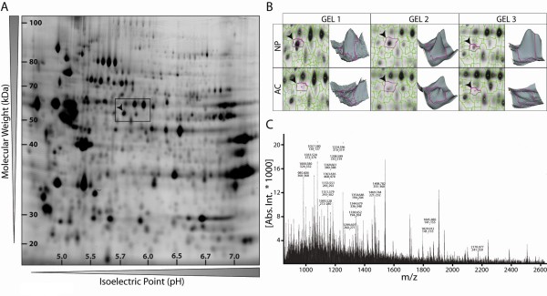
Figure 1.
Proteome map of freshly isolated bovine nucleus pulposus (NP) cells and identification of cytokeratin 8 (CK8) by mass spectrometry. (a) Representative two-dimensional CyDye3-stained gel of total NP cell proteins separated according to their isoelectric point and molecular weight and (b) analysis of the NP and articular chondrocyte (AC) spot patterns by DeCyder software. (c) Mass spectrum of the spot indicated by an arrowhead in (a) and (b) identified by peptide mass fingerprinting as CK8.

