Figure 1.

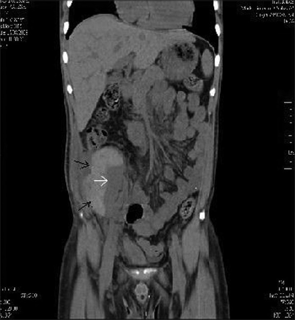
CT of the abdomen-(coronal section) showing sub capsular hematoma (black arrows) compressing the graft kidney (white arrow)

CT of the abdomen-(coronal section) showing sub capsular hematoma (black arrows) compressing the graft kidney (white arrow)