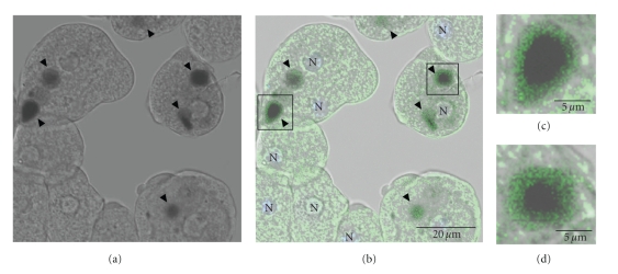
Figure 8.
Cellular localization of EhVps4 protein during erythrophagocytosis. Trophozoites transfected with pEhVps4 were incubated with RBC, treated with diaminobenzidine, anti-rEhVps4 and FITC-labeled secondary antibodies, and analyzed through confocal laser microscopy. (a) Cells observed in phase contrast. (b) Trophozoites observed in the green (FITC) channel and phase contrast. (c) Magnification of diaminobenzidine stained RBC squared in (b). Arrowhead, EhVps4 signal around RBC. N, nuclei.

