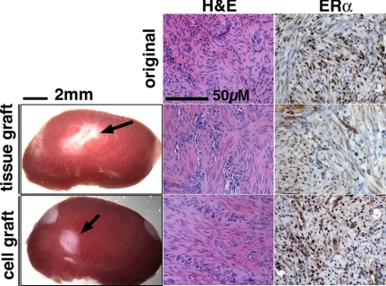
Figure 1.
Gross appearance and histology of myometrial xenografts. Gross appearance of myometrial tissue and cell xenografts (indicated by an arrow) on the host kidney (treated with E2+P4) at 8 wk after grafting. Although the myometrial xenografts did not increase in size with any hormone treatment, both tissue and cell xenografts (with E2 treatment) showed typical histology of myometrium comparable with the original tissue (top panels) in H&E and ERα-IHC staining.

