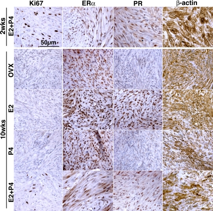
Figure 5.
Histology of UL xenografts subjected to hormone withdrawal. Proliferation activity significantly declined in response to withdrawal of E2 and/or P4 as assessed by Ki67 expression. PR was also down-regulated in response to E2 withdrawal. However, UL xenografts did not show sign of tissue degradation or cell death, and expression of ERα and β-actin was maintained without E2 and/or P4, even after 8 wk. All histology images were taken under a ×20 objective lens and in the same magnification.

