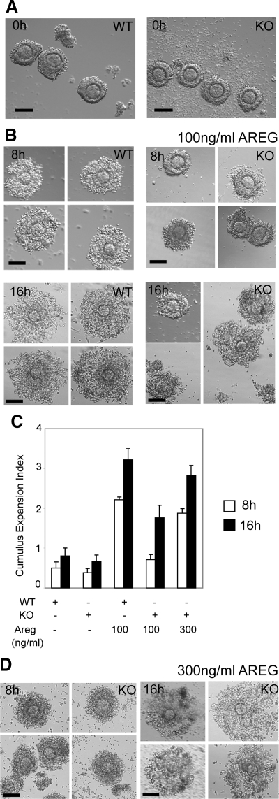
Figure 2.
Restoration of cumulus expansion in RIP140 null COCs. A, COCs released from WT and RIP140 knockout (KO) ovaries after 48-h eCG treatment. Scale bar, 100 μm. B, COCs isolated from WT and RIP140 KO ovaries after 48-h eCG treatment and cultured for 8 and 16 h in the presence of 100 ng/ml AREG. Scale bar, 100 μm. Compared with the WT COCs, the KO COCs showed defects in expansion at 8 h (P < 0.0001) with some COCs showing matrix formation by 16 h. C, Cumulus expansion index scored for COCs. D, COCs isolated from RIP140 KO ovaries after 48-h eCG treatment and cultured for 8 and 16 h in the presence of 300 ng/ml AREG showed improvement in cumulus expansion at 8 h and 16 h. Scale bar, 100 μm. Each treatment was performed with a pool of eight to ten COCs isolated from two to three mice, and three to four independent experiments were performed.

