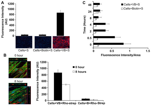
Fig. 4.
(A) Modification of the MSCs with biotinylated lipid vesicles (VB) measured as a function of fluorescent signal after addition of Rhodamine–Streptavidin (S). (B) Confocal image of MSCs with biotinylated lipid vesicle and S immediately after modification (0 h) and 8 h after modification. Green-Actin, Blue-Nucleus, Red-Rhodamine–streptavidin conjugated to biotinylated lipid. Stability of streptavidin conjugated to biotin on cell surface measured immediately and 8 h after modification. (C) Accessibility (and stability) of lipid biotin or adsorbed biotin on the MSC surface measured after addition of S at each time point.

