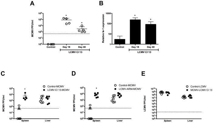
Figure 7. Secondary virus replication in spleen and liver.
Mice uninfected, infected with LCMV Cl 13 at day 18 to 40 pi (A, B and C) or infected with LCMV ARM at day 5 pi (D) were injected with MCMV. In E, uninfected or MCMV-infected mice at day 2.5 pi were challenged with LCMV. Spleens (A–E) and livers (C–E) were collected at day 4 pi. (A, C, D and E) Titers of secondary virus were determined by plaque assay as described in material and methods section. Each symbol represents an individual mouse and dotted line indicate the detection limit of the plaque assay B) MCMV Ie-1 gene was determined by Q-PCR. Results are representative from 2 independent experiments. The mean ± s. d. obtained from 4–6 mice per group is shown. (* dual viral infection compared to single viral infection p<0.01).

