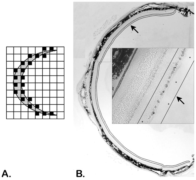
Figure 1.

Uniform random sampling of a tissue section (A) and a retinal cross section (B). The area around the retina is delineated (black line/arrows) with the CAST system and random systematic samples, locations marked by + symbols (inset), are taken throughout the entire delineated area.
