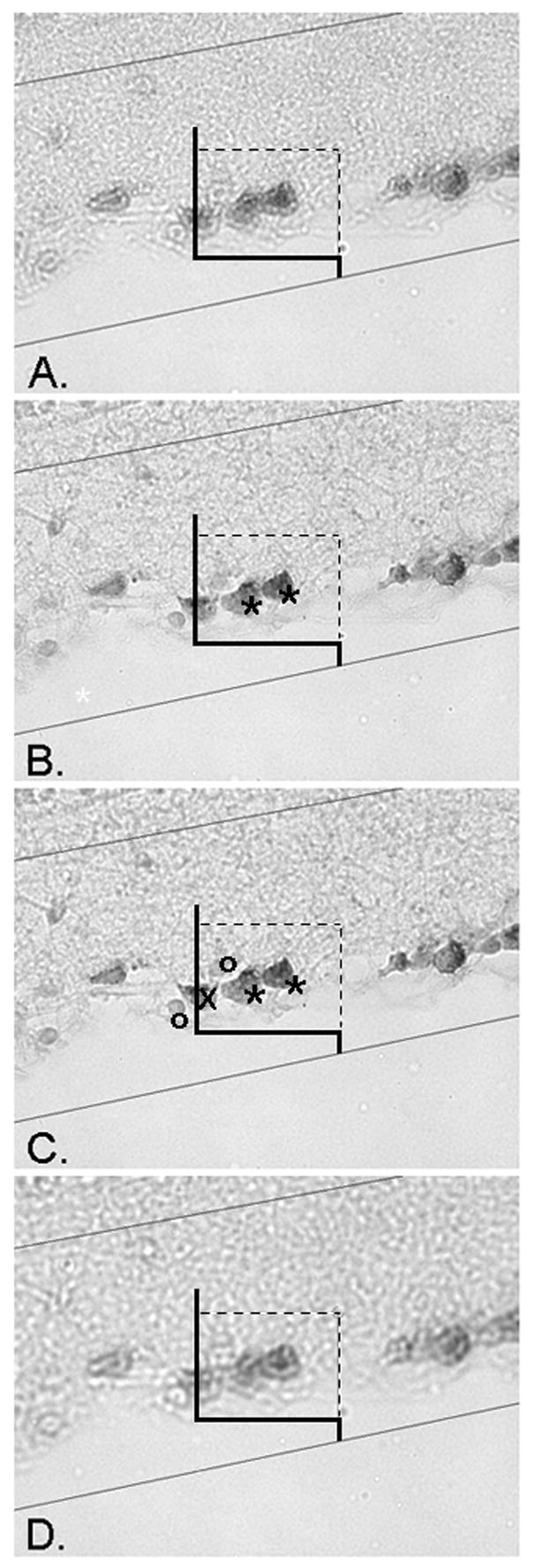
Figure 3.

Application of the optical dissector. The counting frame is applied in a randomly sampled area of the retina, than systematically moved and sampled throughout the entire section (Figure 1). At each sampled position the focal plane of the dissector is moved through the thickness of the section and RGC profiles counted. The upper and lower planes of the section serve as exclusion areas, where profiles are not counted. Micrograph A displays the uppermost plane of the section, when an object first comes into focus. It is important to note that any profiles in focus at this upper plane are not counted. The focal plane is moved through the section from (A) to (D) and all RGCs that come into focus within the frame, obeying the counting frame rules (Figure 2), are counted. In micrograph B, two RGCs are marked with asterisks and counted because they are within the frame, but not in contact with the two forbidden dashed exclusion lines or their extensions. Micrograph C displays the same two counted RGCs further in the focal plane of the section. Note two non-RGC cells marked by circles that are not counted, in addition to the RGCs marked by an X intersecting with the dashed forbidden exclusionary line, and therefore not counted. The plane is focused through the section until the end (D). In this 4 level example, the height of the dissector, h, from level (A) to level (D) is 11 μm.
