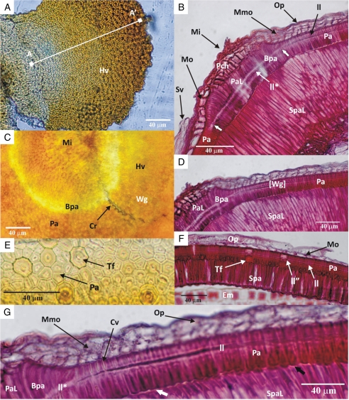
Fig. 6.
Light micrographs of G. carolinianum: (A) top view of hinged valve (the line A–A′ indicates the increasing size of the palisade cells); (B) longitudinal section through micropylar region of a dormant seed (the two short thick white arrows demarcate the widened light line in the micropylar region); (C) periclinal section through the water gap with the hinged valve; (D) longitudinal section of water gap; (E) periclinal section of the palisade layer away from the water gap; (F) longitudinal section of the seed coat away from the water gap; (G) close-up of a longitudinal section of a water gap (the short, thick white or black arrow indicates subpalisade cells with smooth or rough outer periclinal walls, respectively. Abbrevioations: Bpa, bent palisade cells; Cr, crack demarcates the margin of the water gap; Cv, cell lumen; Em, embryo; Hv, hinged valve; ll, light line; ll*, widened light line in the micropylar region; ll'', raised light line in the tanniferous cells; Mi, micropyle; Mmo, multi-layered middle parenchyma cells; Mo, single layer of middle parenchyma cells; Op, outermost polygonal parenchyma cell layer; Pa, palisade cells; PaL, elongated palisade cells of micropylar region; Pch, parenchyma cells of micropyle; Spa, subpalisade cells; SpaL, elongated subpalisade cells of micropylar region; Sv, seed-coat vascular tissue; Tf, tanniferous cells in palisade layer; Wg, water gap open; [Wg], water gap closed.

