Fig. 7.
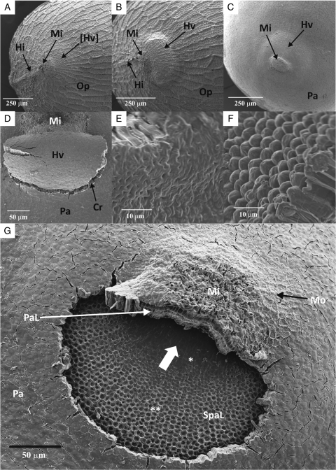
Scanning electron micrographs of Geranium carolinianum seeds: (A) micropylar area of a dormant seed; (B) micropylar area of a non-dormant (heat-treated) seed immersed in water for 10 min; (C) micropylar area of a non-dormant seed without outer permeable cell layers; (D) raised hinged valve of a non-dormant seed without outer permeable cell layers (soaked in water for 10 min); (E) relatively smooth inner periclinal cell walls of water-gap palisade cells near the micropyle; (F) convex-shaped inner periclinal cell walls of water-gap palisade cells near radicle end; (G) water-gap opening of a non-dormant seed without outer permeable cell layers and hinged valve dislodged (soaked in water for 20 min). Abbreviations: Cr, crack demarcates the margin of the water gap; Hi, hilum; Hv, hinged valve (opened); [Hv], hinged valve (closed); Mi, micropyle; Mo, single layer of middle parenchyma cells; Op, outermost polygonal parenchyma cell layer; Pa, palisade cells; PaL, elongated palisade cells of the micropyle; SpaL, elongated subpalisade cells; *, subpalisade cells with smooth outer periclinal cell wall; **, subpalisade cells with concave outer periclinal cell wall. The large white short arrow in (G) indicates the region of initial water uptake.

