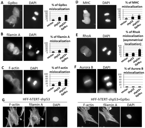
Figure 4. GpIbα-overexpression causes mislocalization of some cytokinesis-related proteins.
(A–E). Left panels: Immunofluorescence revealed that GpIbα, filamin A, F-actin, and MHC were frequently absent and RhoA asymmetrically localized at the cleavage furrow in HFF-shp53+GpIbα cells during cytokinesis. Right panels: The means and standard deviations of the protein mislocalization (n>100 cells per sample). (F) Aurora B localization was not affected by GpIbα overexpression or p53 deletion (n = 100 cells per sample). (G) Interphase localizations of F-actin and filamin A were not affected by GpIbα overexpression. For panels A–E, the p value between HFF-vector and the HFF-shp53+GpIbα cells are each <0.05 by an unpaired two-tailed Student's t-test.

