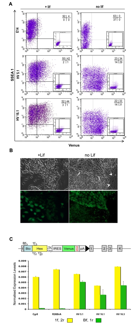
Figure 2. Expression of Venus in a subpopulation of SSEA 1 positive HV cells under self-renewing conditions.
(A) Flow cytometry of two independent HV clones (HV 5.1 and HV 16.1) cultured either under self-renewing conditions or in the absence of LIF show the presence of a subpopulation of cells positive for Venus and/or the ES cell surface marker SSEA-1. Gates for expression of Venus and the presence of SSEA 1 were based on unstained E14 ES cells. Upon the removal of LIF for 3 d, the percentage of cells negative for SSEA 1 increased in both HV clones and the E14 cell line. (B) Fluorescence microscopy of the HV cell line in the presence or absence of LIF. Cultures were differentiated as (A). Note the brighter intensity of Venus in the tightly apposed pavement-like cells in the LIF negative culture (white arrows). Venus expression is absent from giant flat cells (white arrowheads). (C) Expression of the Venus transgene is similar to the low-level expression of the Hex cDNA. RNA was prepared from self-renewing cultures of three HV clones, parental R26BirA cells, and Cgr8 cells. Quantitative PCR analysis was carried out to monitor levels of mRNA derived from both targeted and untargeted alleles of Hex (1f, 2r) or targeted allele only (Bf, 1r). The schematic diagram depicts the different primers used. Values for each primer set used were normalised to the levels of Actin value obtained for each sample.

