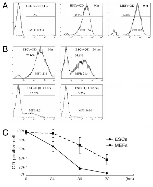
Figure 2.
Quantitative analyses of QD-positive cells by flow cytometry. (A) Representative histograms of QD fluorescence in unlabeled ESCs, labeled ESCs and MEFs right after labeling. (B) Histograms of QD-labeling in ESCs during cell culture. (C) Dynamic changes of QD-labeling in ESCs and MEFs were followed up to 72 hours by flow cytometry. MFI: mean fluorescence intensity of the whole population.

