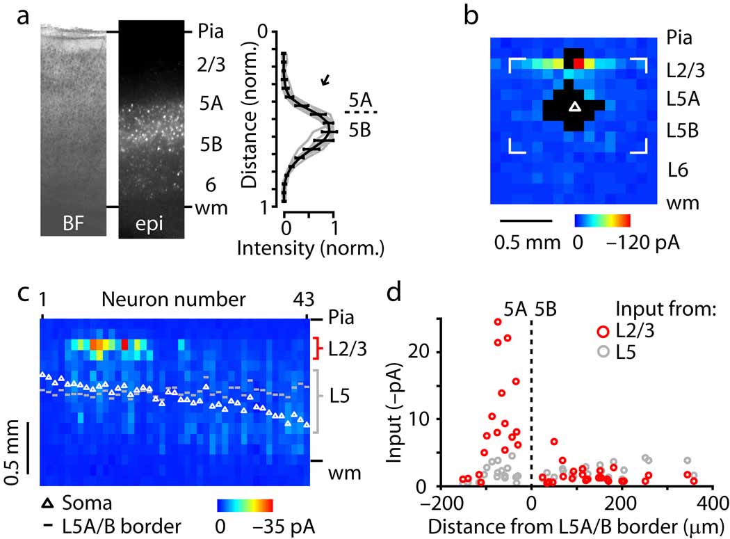
Fig. 2.
Sub-layer specific circuits of crossed corticostriatal projection neurons. (a) Laminar distribution of fluorescently labeled neurons. (b) Example of corticostriatal neuron input map. (c) Side view of group of input maps (n = 43 corticostriatal neurons). (d) Layer 2/3 (red) and 5 (gray) input as a function of the distance of the soma from the layer 5A/B border, along the radial axis of the cortex (pia is leftward and white matter is rightward).

